Can a Repaired Hernia Rupture Again
Introduction
Umbilical hernias are nigh common in women than men. Pregnancy may cause an umbilical hernia, or render a preexisting 1 credible, because of progressively increasing intra-abdominal pressure level. Hernia symptoms present in the second trimester in nearly patients. A hernia may exist diagnosed during first, 2d, or tertiary pregnancies (ane). The incidence of an umbilical hernia in significant women has been reported to be as low as 0.08% in a very recent big series (ii). However, it is possible to run across complicated cases, like a total-term pregnancy in umbilical hernia (3), peritonitis due to peel ulceration (iv), or incarcerated significant uterus within the hernia rims (5).
A surgical algorithm for a significant adult female with a hernia is not clear to engagement, but newer and better scientific data has been cumulated (1, two, half dozen). There is no consensus about the timing of surgery for an umbilical hernia in a woman who is already significant or planning a pregnancy. In fact, these two types of cases should be taken into consideration separately. Augustin and Majerovic recommended that hernias that are symptomless or have minimal symptoms—including slight discomfort or pain—should be examined regularly and cured electively later delivery and uterine involution (7). Recently, it has been shown that watchful waiting, even up to 5 years, appears to be a safe strategy for ventral hernias in the adult population (8).
It seems to be better and more understandable to stratify the cases into several scenarios regarding the relationship betwixt umbilical hernia and pregnancy. In fact, discussing the issue on a example-by-case basis may be the best arroyo.
Umbilical Hernia in Women Planning for a Pregnancy
In this situation, we have several concerns.
• Should nosotros repair the hernia earlier pregnancy?
• Which repair technique should be used?
• Tin the repair remain intact during pregnancy?
• Tin can the repair cause pain and discomfort during pregnancy?
• How long should the interval betwixt the hernia repair and the pregnancy or nascence be?
• What complications can happen during pregnancy if we leave the hernia unrepaired?
When the hernia is incarcerated or strangulated at the time of diagnosis, an emergency repair is inevitable. If the hernia is not complicated, but symptomatic, an elective repair should be proposed. A symptom may be pain or a large bulging. When the patient has a modest and asymptomatic hernia, information technology may exist better to postpone the repair until afterwards she gives birth. Fortunately, most of the cases nosotros encounter are in this group. During pregnancy, the enlarged uterus pushes the intestinal loops to superior and posterior parts of the intestinal crenel. The size and pushing force of the uterus during the first trimester does non seem enough to push the intestines into a pocket-size umbilical opening. The uterus reaches the level of the umbilicus at well-nigh the 20th–22nd week (9, ten). Thereafter, no close contiguity betwixt umbilical hernia defect and intestinal segments be (Figure ane). If an incarceration occurs during this time, there is less concern most the surgical intervention, considering an performance in the outset or 2d trimester would not carry high risks for preterm labor or other adverse effects (eleven).
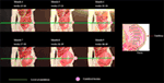
Figure 1. Changes in the size of the uterus and its relation to the umbilicus past the weeks of pregnancy.
A proper repair technique for an umbilical hernia in a woman planning a pregnancy is also a question. Information technology has been shown that mesh repairs provide ameliorate outcomes than suture repairs (12). Repairing with just sutures may bring a recurrence during pregnancy (6). Lappen et al. reported that pregnancy caused an increased hazard of abdominal hernia recurrence. This information should exist given to the patients who are planning an constituent hernia repair before a subsequent gestation (13). As the uterus enlarges and intra-abdominal pressure rises, fifty-fifty mesh repairs will non make a significant woman immune to hernia recurrence. In cyclopedia, Oma et al. reported that pregnancy after umbilical hernia repair was independently associated with ventral hernia recurrence and mesh utilize could non lower the run a risk of recurrence (fourteen). A repair with mesh may restrict the flexibility of the abdominal wall (15) and may cause pain during a subsequent pregnancy (16).
Unfortunately, there is no substantial evidence about the adequate interval between hernia repair and pregnancy or birth. Surgeons usually propose their patients that a pregnancy is not immune until after the offset twelvemonth of the surgical repair. All the same, no clinical or experimental studies exist on this specific case. At that place is no consensus on if this ane-twelvemonth interval ends at the starting time of the pregnancy or at the fourth dimension of birth. It can only be said that an early on pregnancy may crusade recurrence.
Every hernia carries a chance of incarceration and strangulation. Therefore, patients with an umbilical hernia and planning a gestation should be instructed about this take chances. No ane can predict which hernias will become complicated or when this volition occur. However, every surgeon can tell his or her patient what the malicious furnishings of an incarcerated or strangulated hernia are on the mother and the baby. An emergency repair, specially during the first or third trimester, will bring the burden of anesthesia and surgical trauma. It should be recommended that patients with large hernias, including intestinal loops, umbilical hernias with a suspicious history of incarceration, and recurrent umbilical hernias previously repaired with a mesh undergo a definitive repair before planning a pregnancy (Effigy ii).
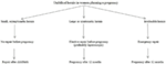
Figure 2. Surgical strategy for umbilical hernia in women planning a pregnancy.
Umbilical Hernia Diagnosed during Pregnancy
Over again, there is no solid recommendation for this type of case. Unfortunately, no randomized controlled trial or prospective analysis about hernia repairs in pregnancy existed in the literature (vi). However, a pocket-size asymptomatic or minimally symptomatic umbilical hernia diagnosed in the early stage of a pregnancy tin can be managed like a hernia in women planning to get pregnant (Figure 3). Symptomatic umbilical hernias can sally in every trimester of pregnancy, and they may become incarcerated or strangulated during pregnancy, although the verbal rates of these complications have never been reported. Haskins et al. reviewed the American College of Surgeons National Surgical Quality Comeback Programme and plant that 126 pregnant women were operated on for umbilical hernia repair in a 10-yr period (17). 90-5 percent of the repairs performed with open technique. Incarceration or strangulation existed in half of the cases. Surgery was achieved with minimal 30-twenty-four hours morbidity for the mother and no fetal loss, even in cases of emergencies. Buch diagnosed v female person patients with umbilical hernias occurring during pregnancy at the Mount Sinai Medical Eye from September 2004 to July 2006 (one). All patients presented with symptoms in the second trimester with reducible hernias. None of them adult incarceration until an open repair following delivery. This finding supports watchful waiting approach during pregnancy (1).
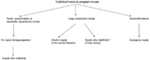
Effigy 3. Surgical strategy for an umbilical hernia diagnosed during pregnancy.
30-i papers, including twenty-three case reports, were found in a recent literature search by Jensen et al. (half dozen). Apart from the above cases mentioned by Haskins and Buch (i, 17), 7 patients with an umbilical hernia underwent emergency repair during pregnancy. Suture repair was used in all cases, merely one. Wai et al. from Yale Academy, reported the unique case, describing an intraperitoneal mesh repair for an irreducible umbilical hernia in a woman in the second trimester (eighteen). In Jensen et al.'southward literature review, no postoperative complications were recorded (half-dozen). This included Ahmed's emergency repair case with spontaneous rupture in a young, multiparous woman in 28th week of pregnancy (19). In that case, there was skin ulceration due to pressure, and the uterus was completely in the hernia sac with gangrenous intestinal loops of approximately 75 cm. The hernia defect was closed with a suture and the patient gave birth uneventfully vi weeks later (xix).
Oma et al. published the almost recent series (ii). In this series, 17 pregnant women with an umbilical hernia were recorded within 20,714 pregnancies in a single institution. There were v pregnant patients with an umbilical hernia. Ii women noticed the hernia during previous pregnancies, one patient in the present gestation, and the other two at 5th week of pregnancy. All patients completed their pregnancies with no hernia complication.
Cesarean Section (C-Section) and Simultaneous Hernia Repair
Hernia repair during C-section is a mutual surgical approach. Withal, these simultaneous surgeries were not well-documented until the 2000s. In 2004, Ochsenbein-Kölble et al. reported the first case series of C-sections and simultaneous inguinal or umbilical hernia repairs (20). Three patients were offered and underwent combined surgery with their informed consent. In one of them, the sign for C-section was the presence of the umbilical hernia itself. The duration of surgery was longer in cases with inguinal hernia repair, only non umbilical hernia repair versus C-department lonely. However, Ghnnam et al. reported that the simultaneous umbilical hernia repair and cesarean needed more than time than just a cesarean (21). They compared 48 patients, who underwent cesarean delivery along with paraumbilical hernia repair versus 100 patients undergoing a C-section. Inpatient periods were like. Only ii patients complained of pain at the bellybutton. The control group needed significantly fewer analgesics. Combined surgery was preferred by all patients. One hernia recurred (2.8%), following suture repair within ii years (21). Mesh repairs were complimentary of recurrence.
Gabriele et al. reported on 28 pregnant women with an inguinal or an umbilical hernia. These patients who underwent simultaneous C-department and hernia repair were compared with 100 patients who but underwent a C-section (22). Combined surgeries took more than time for both an umbilical and inguinal hernia than C-section solitary. Surgeries were uneventful, and no recurrence adult. The authors ended that combined surgery is safe and avoids readmissions. Also, Jensen et al. came to a solid determination after their literature search that combined hernia repair and C-department is the optimal therapeutic choice (half dozen).
Steinemann et al. recently published a retrospective cohort–control study (23). Fourteen patients underwent suture repair of umbilical hernia during C-section by using different techniques. External umbilical hernia repair with suture was used in vii cases via a paraumbilical semilunar skin incision later on the closure of the Pfannenstiel incision. Internal umbilical hernia repair with suture was used in the other seven patients. Internal suturing required less time than external suturing. Both approaches lengthen the time in operation compared to the command group. Unfortunately, two recurrences were revealed by ultrasonography in each repair subgroup (28%). The authors recommended mesh repairs in these cases (23).
Interestingly, no patient underwent combined surgery in Haskins et al.'s nigh recent review (17). However, the reason for the absenteeism of whatsoever instance with simultaneous C-section and hernia repair are non explained in the paper.
Hernia Repair after Childbirth following an Interval
Some significant women with an umbilical hernia do not undergo simultaneous hernia repair at the time of C-section. The reason for that may exist a patient'due south or surgeon's option.
Oma et al. followed eight women with an umbilical hernia and no surgical intervention throughout their pregnancy. The umbilical hernia persisted in all these patients who had a clinical re-evaluation postpartum and no spontaneous disappearance of the hernia was recorded. Elective umbilical hernia repairs were washed in five patients within 5 months to 3 years after delivery (2).
Buch et al. reported five cases that underwent hernia repair in the postpartum period. The patients underwent surgery at postpartum for 8–52 weeks. No complications or recurrence were recorded in postoperative follow-up for 2–34 weeks. Ii out of v women conceived over again after hernia repair. The authors concluded that pregnant patients presenting with reducible groin or umbilical hernias during pregnancy can safely be managed non-operatively during their pregnancy and undergo surgical repair in the postpartum flow (1).
Combined surgery may not increment the risk of local and systemic complication (20), however, there are nonetheless other concerns about simultaneous surgery. Apart from maternal and fetal health, there are bug regarding the quality and durability of the hernia repair. What would be the advantages of surgical repair in the postpartum menstruation rather than during C-section? In other words, could a concomitant repair during C-section be less reliable? Let the states accept a look at potential hazards of repair during C-department.
Changes in Muscles and Fascial Structures during Pregnancy
The gross structure of rectus abdominis muscle is altered during pregnancy. Significant increases happen in muscle length, separation, and angles of insertions as the pregnancy progressed (24). The functional ability of the abdominal muscles is also altered, and the ability to stabilize the pelvis is decreased. For all intestinal exercises, upper rectus abdominis relative integrated electromyography (EMG) increased while external oblique and lower rectus abdominis relative integrated EMG decreased. Relative EMG for all tested muscles returned to levels seen at 18 weeks and 26 gestations past 18 weeks post-birth. Functional changes plant in the rectus abdominis and external and internal obliques. During the firsthand post-birth menses, separation of the rectus abdominis was resolved by four weeks post-birth and abdominal muscle inter-relationships returned to early on pregnancy levels by 8 weeks mail-birth. Nevertheless, the ability to stabilize the pelvis remained low at eight weeks post-nascency. This sustained decrement in the power to stabilize the pelvis at eight weeks post-birth may reflect the poor resolution of abdominal muscle length increases due to pregnancy (24).
In fact, the significant of the alterations in intestinal muscle groups for the fate of an umbilical hernia repair is obscure. Whether the changes increase or decrease, hernia recurrence rates is unknown to surgeons. However, the abdominal muscles during pregnancy differ from usual. It may exist improve to await for a while to let the muscles return to their normal anatomy and function earlier repairing the umbilical hernia. However, at that place is no recommendation in the literature for the exact fourth dimension to await for a repair.
Relaxin. Is information technology ImWportant?
Another issue that may touch on the fate of hernia repair in a significant or early on postpartum woman is hormonal changes during gestation. Relaxin is a peptide hormone in the insulin family unit, secreted past the corpus luteum (25). It is besides released from the placenta during pregnancy. It relaxes pelvic ligaments and softens and widens the neck. Relaxin reduces extracellular matrix (ECM) synthesis and induces collagen degradation (26). In a report on rats, relaxin caused a significant reduction in tissue collagen content (27). Relaxin limited collagen production, while stimulating increased collagen degradation (28). Also, Naqvi et al. documented relaxin'southward degradative effects on joint fibrocartilaginous tissue with matrix degradation by metalloproteinases (MMPs) (29).
Collagen, ECM, and MMPs have important implications for hernia formation. Collagen is the near abundant ECM protein. Collagenase, a member of the MMP family, is the master enzyme in collagen deposition (30).
Considering the studies on the human relationship between collagen, ECM, and MMPs, we can think any endogenous or exogenous substance that affects these mechanisms may crusade recurrence later hernia repair, especially following suture repairs. Therefore, we can say at that place may be a risk of recurrence when the repair is washed and the relaxin level is high. Although in that location is no prove for this supposition, there are interesting reports in the literature. It has been reported that a higher expression of relaxin receptors within the muscles of the pelvic diaphragm in dogs with a perineal hernia. This may suggest that relaxin plays a function in the pathogenesis of this blazon of hernia by causing muscular cloudburst (31). Relaxin may also be a factor in perineal hernia formation with connective tissue degeneration in dogs (32). In homo beings, there is only 1 report on the relation between relaxin and abdominal hernias (33). In this written report, all the children born in Malmö, Sweden in a 5-year catamenia were checked for built dislocation of the hip (CDH) and for an inguinal hernia. Hernia was diagnosed five times more frequently in girls with CDH than girls without, and iii times in boys with CDH than boys without. The authors stated that relaxin could stimulate collagenase, induce structural changes in the connective tissue, and cause evolution of both CDH and the hernia (33). This paper was published in 1988 and no further information on the subject has been nerveless since.
Would Lifting and Carrying Infant Create a Brunt on the Repair?
Surgeons generally put patients on a weight lifting restriction after hernia repairs. Even mesh repairs are vulnerable to rises in intra-intestinal pressure in the early postoperative period. Biomechanical studies have revealed that the tensile forcefulness provided by tissue ingrowth into the mesh reaches approximately eighty% after just vi weeks (34). Although in that location is no consensus on weight lifting restriction after hernia repairs, surgeons exercise not desire their patients to lift whatever weight for the first 2 weeks. Moderate lifting (<10 kg) is allowed after 2–4 weeks. Patients are advised to lift over 10 kg only after eight weeks (35). In fact, carrying and lifting a baby would stay within the limits of the advice. Still, a woman who does not accept a baby and undergoes umbilical hernia repair would be on a weightlifting restriction for a much longer time.
Although umbilical hernia repair tin can be performed after childbirth, at that place is no need for surgery on modest asymptomatic hernias in the early postpartum catamenia. A 1-year interval can give the patient a very smooth convalescence, including hormonal stabilization and return to normal body weight. Surgery tin exist postponed for a longer time, even after another pregnancy, if the patient would similar to have more children.
Significance of the Concomitant Diastasis Recti (DR)
Diastasis recti is the midline separation of the rectus abdominis muscles. It is an damage, just not a truthful hernia, and does non acquit a risk for incarceration. There is a positive correlation betwixt parity and DR (36). The prevalence during pregnancy is about xxx–lxx%. The normal width of the linea alba is fifteen mm at the level of xiphoid, 22 mm at the level of 3 cm cranial to the umbilicus, and 16 mm at the level of 3 cm caudal to the umbilicus in nulliparous women (37). Mechanical forces and hormonal changes during pregnancy may play a role in the etiology.
The nearly frequent localization is in the periumbilical region and persistence postpartum is plant in almost lx% of cases (38). Liaw et al. reported that diastasis may persist in the postpartum menstruum and the abdominal muscle office improved, but did not return to normal, even subsequently half dozen months (39). Sperstad et al. followed 300 first-time pregnant women from pregnancy until 12 months postpartum. They reported that DR existed in 33.one, sixty.0, 45.4, and 32.half dozen% of the women at 21 weeks of pregnancy, and at 6 weeks, 6 months, and 12 months following delivery, respectively (40). This study revealed that the adventure for DR was twofold college in women reporting heavy lifting xx times a week or more than in women reporting less weight lifting. The authors did not describe the heavy lifting in the text, only nosotros can assume that a postpartum woman lifts her baby many times a week. The weight of a babe is about 8 kg at half-dozen months and 10 kg at 12 months (41). These weights are plenty to raise intra-abdominal pressure level every bit high as a Valsalva maneuver does (35).
Although RD is not a hernia, it may cause recurrence as a larger hernia following umbilical hernia repairs. In umbilical hernia repairs with sutures, the bites pass through a weak rectus sheet at the region of diastasis. This may cause tears and create button pigsty defects, consequently resulting in recurrence. Köhler et al. evaluated 231 suture repairs for small primary umbilical or epigastric hernias (42). Hernia defects were smaller than 2 cm. Patients with rectus diastasis developed hernia recurrence at a significantly increased rate. The authors hypothesized that thin and stretched rectus sheath is a risk factor for recurrence. They recommended mesh repair for umbilical hernia patients with rectus diastasis. Although Emanuelsson et al.'s recent prospective randomized study showed that two-row suture plication with delayed absorbable fabric provided similarly good results with retromuscular lightweight polypropylene mesh without an increase in recurrence rate in treatment of RD (43), mesh apply remains a ameliorate option for patients with concomitant umbilical hernia and RD (42). In addition, one can assume that a recurrence withal may develop from the sites of mesh fixation if in that location is a vulnerable linea alba due to RD. Therefore, it is ameliorate to utilise no fixation in case of potent restoration of the line alba or to use an autraumatic mesh fixation like glues (e.1000., fibrin) (44) or a self-gripping mesh in retromuscular mesh repairs (45).
Determination
There is no consensus about the timing of surgery for an umbilical hernia in a woman who is already pregnant or planning a pregnancy. If the hernia is incarcerated or strangulated at the time of diagnosis, an emergency repair is inevitable. If the hernia is not complicated, just symptomatic, an elective repair should be proposed. If the hernia is repaired by suture, the risk of recurrence is high during pregnancy. Repair with a mesh may restrict the flexibility of the intestinal wall and may cause pain during a subsequent pregnancy. When the patient has a small and asymptomatic hernia, it may exist improve to postpone the repair until she gives birth.
Umbilical hernia repair during pregnancy tin can be performed with minimal morbidity to the mother and no fetal loss even in emergency cases. If a small hernia becomes larger and symptomatic, the second trimester is a proper period for surgery. Umbilical hernias can be repaired post-obit childbirth or at the time of C-department. Patient satisfaction is high for combined C-section and hernia repair. Nevertheless, a high recurrence rate is expected.
Elective repair after childbirth is well-documented. It is possible as early as the postpartum at 8 weeks. In that location is no need for surgery for small asymptomatic hernias in the early postpartum menses. A i-year interval tin can give the patient a very smooth convalescence, including hormonal stabilization and return to normal torso weight. Surgery tin be postponed for a longer time, even after some other pregnancy, if the patient would like to have more children.
Diastasis recti are very frequent during pregnancy. It may persist in the postpartum flow. Patients with rectus diastasis may develop umbilical hernia recurrence after repair. This take a chance is especially loftier post-obit suture repairs. Mesh repairs should be considered in this state of affairs (Table 1).

Table 1. Pros and cons for specific conditions in the relation of umbilical hernia and pregnancy.
Author Contributions
The author confirms being the sole contributor of this work and canonical it for publication.
Conflict of Interest Statement
The author declares that the enquiry was conducted in the absence of any commercial or financial relationships that could be construed as a potential conflict of interest.
References
2. Oma E, Bay-Nielsen Yard, Jensen KK, Jorgensen LN, Pinborg A, Bisgaard T. Primary ventral or groin hernia in pregnancy: a cohort study of 20,714 women. Hernia (2017) 21(3):335–9. doi:10.1007/s10029-017-1618-7
PubMed Abstract | CrossRef Full Text | Google Scholar
5. Shaw WG, Nichols EE. Abdominal pregnancy incarcerated into an umbilical hernia. Instance report. Am J Obstet Gynecol (1962) one(84):72–v. doi:10.1016/0002-9378(62)90675-0
CrossRef Full Text | Google Scholar
8. Kokotovic D, Sjølander H, Gögenur I, Helgstrand F. Watchful waiting equally a handling strategy for patients with a ventral hernia appears to exist safe. Hernia (2016) 20(2):281–vii. doi:ten.1007/s10029-016-1464-z
PubMed Abstract | CrossRef Full Text | Google Scholar
9. Lowdermilk DL. Beefcake and physiology of pregnancy. 11th ed. In: Lowdermilk DL, Perry SE, Cashion MC, Alden KR, editors. Maternity and Women'southward Wellness Intendance – E-Book. St. Louis: Elsevier (2004). p. 283–300.
Google Scholar
10. Merritt TA, Sounders BA, Gluck L. Antenatal assessment of fetal maturity. second ed. In: Aladjem S, Brown AK, Sureau C, editors. Clinical Perinatology. St. Louis: Mosby (1980). 214 p.
Google Scholar
13. Lappen JR, Sheyn D, Hackney DN. Does pregnancy increase the risk of abdominal hernia recurrence after prepregnancy surgical repair? Am J Obstet Gynecol (2016) 215(3):390.e1–5. doi:10.1016/j.ajog.2016.05.003
PubMed Abstract | CrossRef Full Text | Google Scholar
14. Oma Eastward, Jensen KK, Jorgensen LN. Recurrent umbilical or epigastric hernia during and later pregnancy: a nationwide cohort written report. Surgery (2016) 159(6):1677–83. doi:x.1016/j.surg.2015.12.025
CrossRef Full Text | Google Scholar
xv. Junge G, Klinge U, Prescher A, Giboni P, Niewiera Chiliad, Schumpelick 5. Elasticity of the anterior abdominal wall and touch on for reparation of incisional hernias using mesh implants. Hernia (2001) five(3):113–viii. doi:ten.1007/s100290100019
PubMed Abstract | CrossRef Full Text | Google Scholar
xvi. Schoenmaeckers E, Stirler 5, Raymakers J, Rakic S. Pregnancy post-obit laparoscopic mesh repair of ventral abdominal wall hernia. JSLS (2012) xvi(one):85–8. doi:ten.4293/108680812X13291597716104
PubMed Abstract | CrossRef Full Text | Google Scholar
17. Haskins IN, Rosen MJ, Prabhu AS, Amdur RL, Rosenblatt S, Brody F, et al. Umbilical hernia repair in pregnant patients: review of the American College of Surgeons National Surgical Quality Improvement Program. Hernia (2017) 21(5):767–70. doi:10.1007/s10029-017-1633-8
PubMed Abstract | CrossRef Full Text | Google Scholar
18. Wai PY, Ruby JA, Davis KA, Roberts AC, Roberts KE. Laparoscopic ventral hernia repair during pregnancy. Hernia (2009) 13(5):559–63. doi:10.1007/s10029-009-0476-3
CrossRef Full Text | Google Scholar
20. Ochsenbein-Kölble N, Demartines N, Ochsenbein-Imhof N, Zimmermann R. Cesarean section and simultaneous hernia repair. Arch Surg (2004) 139(viii):893–five. doi:10.1001/archsurg.139.8.893
PubMed Abstruse | CrossRef Full Text | Google Scholar
21. Ghnnam WM, Helal AS, Fawzy M, Ragab A, Shalaby H, Elrefaay East. Paraumbilical hernia repair during cesarean delivery. Ann Saudi Med (2009) 29(2):115–8. doi:10.4103/0256-4947.51798
PubMed Abstract | CrossRef Full Text | Google Scholar
23. Steinemann DC, Limani P, Ochsenbein N, Krähenmann F, Clavien PA, Zimmermann R, et al. Suture repair of umbilical hernia during caesarean department: a example-command written report. Hernia (2013) 17(4):521–6. doi:10.1007/s10029-013-1087-6
PubMed Abstract | CrossRef Total Text | Google Scholar
24. Gilleard WL. The Structure and Office of the Abdominal Muscles during Pregnancy and the Firsthand Mail-Birth Catamenia. Master thesis, Wollongong, Australia: Academy of Wollongong (1992).
Google Scholar
26. Samuel CS, Lekgabe ED, Mookerjee I. The effects of relaxin on extracellular matrix remodeling in health and fibrotic disease. Adv Exp Med Biol (2007) 612:88–103. doi:10.1007/978-0-387-74672-2_7
PubMed Abstruse | CrossRef Total Text | Google Scholar
27. Samuel CS, Coghlan JP, Bateman JF. Effects of relaxin, pregnancy and parturition on collagen metabolism in the rat pubic symphysis. J Endocrinol (1998) 159(1):117–25. doi:x.1677/joe.0.1590117
PubMed Abstruse | CrossRef Full Text | Google Scholar
29. Naqvi T, Duong TT, Hashem G, Shiga M, Zhang Q, Kapila S. Relaxin'due south induction of metalloproteinases is associated with the loss of collagen and glycosaminoglycans in synovial joint fibrocartilaginous explants. Arthritis Res Ther (2005) 7(1):R1–11. doi:x.1186/ar1451
PubMed Abstract | CrossRef Full Text | Google Scholar
31. Merchav R, Feuermann Y, Shamay A, Ranen E, Stein U, Johnston DE, et al. Expression of relaxin receptor LRG7, canine relaxin, and relaxin-like gene in the pelvic diaphragm musculature of dogs with and without perineal hernia. Vet Surg (2005) 34(five):476–81. doi:10.1111/j.1532-950X.2005.00072.ten
PubMed Abstract | CrossRef Full Text | Google Scholar
32. Niebauer GW, Shibly S, Seltenhammer Grand, Pirker A, Brandt S. Relaxin of prostatic origin might be linked to perineal hernia formation in dogs. Ann N Y Acad Sci (2005) 1041:415–22. doi:10.1196/annals.1282.062
PubMed Abstract | CrossRef Full Text | Google Scholar
33. Udén A, Lindhagen T. Inguinal hernia in patients with congenital dislocation of the hip. A sign of general connective tissue disorder. Acta Orthop Scand (1988) 59(half-dozen):667–8. doi:10.3109/17453678809149421
PubMed Abstract | CrossRef Total Text | Google Scholar
34. Majercik S, Tsikitis V, Iannitti DA. Strength of tissue attachment to mesh subsequently ventral hernia repair with synthetic composite mesh in a porcine model. Surg Endosc (2006) xx:1671–4. doi:ten.1007/s00464-005-0660-1
PubMed Abstract | CrossRef Total Text | Google Scholar
35. Forbes J, Fry N, Hwang H, Karimuddin AA. Timing of return to work after hernia repair: recommendations based on a literature review. B C Med J (2012) 54(vii):341–5.
Google Scholar
36. Turan V, Colluoglu C, Turkyilmaz Due east, Korucuoglu U. Prevalence of diastasis recti abdominis in the population of young multiparous adults in Turkey. Ginekol Pol (2011) 82(11):817–21.
PubMed Abstract | Google Scholar
37. Beer GM, Schuster A, Seifert B, Manestar M, Mihic-Probst D, Weber SA. The normal width of the linea alba in nulliparous women. Clin Anat (2009) 22(6):706–11. doi:ten.1002/ca.20836
PubMed Abstruse | CrossRef Total Text | Google Scholar
38. Kimmich N, Haslinger C, Kreft K, Zimmermann R. [Diastasis recti abdominis and pregnancy]. Praxis (Bern 1994) (2015) 104(15):803–6. doi:10.1024/1661-8157/a002075
CrossRef Total Text | Google Scholar
39. Liaw LJ, Hsu MJ, Liao CF, Liu MF, Hsu AT. The relationships between inter-recti distance measured by ultrasound imaging and abdominal muscle functionin postpartum women: a half-dozen-calendar month follow-upwardly study. J Orthop Sports Phys Ther (2011) 41(half-dozen):435–43. doi:x.2519/jospt.2011.3507
CrossRef Full Text | Google Scholar
40. Sperstad JB, Tennfjord MK, Hilde G, Ellström-Engh One thousand, Bø Grand. Diastasis recti abdominis during pregnancy and 12 months after childbirth: prevalence, risk factors and report of lumbopelvic pain. Br J Sports Med (2016) 50(17):1092–6. doi:10.1136/bjsports-2016-096065
CrossRef Full Text | Google Scholar
41. World Wellness Organization, Department of Nutrition for Health and Evolution. WHO Kid Growth Standards 1 Yr 2 Years 3 Years 4 Years five Years Length/Pinnacle for Historic period, Weight for Age, Weight for Length, Weight for Height and Trunk Mass Index for Historic period: Methods and Evolution. (2006). Available from: http://www.who.int/childgrowth/standards/technical_report
Google Scholar
42. Köhler G, Luketina RR, Emmanuel Grand. Sutured repair of primary pocket-size umbilical and epigastric hernias: concomitant rectus diastasis is a pregnant take chances cistron for recurrence. World J Surg (2015) 39(1):121–6; discussion 127. doi:10.1007/s00268-014-2765-y
PubMed Abstract | CrossRef Full Text | Google Scholar
43. Emanuelsson P, Gunnarsson U, Dahlstrand U, Strigård G, Stark B. Operative correction of abdominal rectus diastasis (ARD) reduces hurting and improves abdominal wall musculus strength: a randomized, prospective trial comparing retromuscular mesh repair to double-row, self-retaining sutures. Surgery (2016) 160(5):1367–75. doi:x.1016/j.surg.2016.05.035
PubMed Abstract | CrossRef Full Text | Google Scholar
44. Canziani M, Frattini F, Cavalli Thou, Agrusti Southward, Somalvico F, Campanelli K. Sutureless mesh fibrin gum incisional hernia repair. Hernia (2009) thirteen(6):625–9. doi:ten.1007/s10029-009-0555-5
PubMed Abstruse | CrossRef Full Text | Google Scholar
45. Privett BJ, Ghusn M. Proposed technique for open repair of a pocket-sized umbilical hernia and rectus divarication with self-gripping mesh. Hernia (2016) 20(4):527–30. doi:x.1007/s10029-016-1470-1
PubMed Abstract | CrossRef Full Text | Google Scholar
Source: https://www.frontiersin.org/articles/10.3389/fsurg.2018.00001/full
إرسال تعليق for "Can a Repaired Hernia Rupture Again"